Tail of Spence: A Comprehensive Guide to Breast Anatomy and Clinical Significance
The tail of Spence, also known as the axillary process, is an extension of the breast tissue that extends into the axilla (armpit). This seemingly small anatomical feature plays a significant role in breast health and can be a site for various breast-related conditions. This comprehensive guide will delve into the anatomy, clinical significance, and potential issues related to the tail of Spence, providing you with a complete understanding of this important part of the female breast.
This article provides an in-depth look at the tail of Spence, offering a unique perspective on its anatomy, clinical importance, and potential conditions. We will explore its relevance in breast cancer detection, benign breast changes, and surgical considerations. Whether you are a medical professional, a student, or simply interested in learning more about breast health, this guide aims to provide you with valuable insights and practical knowledge.
## Deep Dive into the Tail of Spence: Anatomy, Significance, and Nuances
The tail of Spence is a crucial anatomical extension of the mammary gland that reaches into the axillary region. It’s not merely a superficial feature; it’s a functional part of the breast tissue, containing glandular tissue, ducts, and fat. Understanding its anatomy is essential for accurate clinical assessment and diagnosis of breast-related conditions.
### Comprehensive Definition, Scope, & Nuances
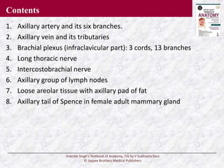
The tail of Spence represents the upper outer quadrant of the breast extending into the axilla. It’s named after James Spence, a Scottish surgeon who described this anatomical feature. Unlike the main breast tissue, the tail of Spence is less defined and can vary in size and shape among individuals. Its proximity to the axillary lymph nodes makes it a significant area for breast cancer metastasis.
### Core Concepts & Advanced Principles
The tail of Spence is composed of the same tissues as the rest of the breast: glandular tissue responsible for milk production, ducts that transport milk to the nipple, and fatty tissue that provides support and volume. The glandular tissue in the tail of Spence is responsive to hormonal changes, just like the rest of the breast, which can lead to cyclical changes and discomfort during menstruation. Its connection to the axillary lymph nodes is a critical consideration in breast cancer staging and treatment planning.
### Importance & Current Relevance
The tail of Spence is clinically relevant because it is a common site for breast cancer development. Due to its location in the axilla, tumors in this area may be mistaken for other conditions, leading to delayed diagnosis. Moreover, the tail of Spence plays a crucial role in breast cancer staging, as the presence of cancer cells in the axillary lymph nodes indicates the extent of the disease. Recent studies indicate that tumors located in the tail of Spence may have different biological characteristics compared to tumors in other parts of the breast.
## Mammography: A Key Tool for Examining the Tail of Spence
Mammography is an X-ray imaging technique used to screen for and diagnose breast cancer. It is one of the primary methods for examining the tail of Spence due to its ability to visualize the entire breast tissue, including the axillary extension. Modern mammography techniques, such as digital mammography and tomosynthesis (3D mammography), provide detailed images of the breast, enhancing the detection of subtle abnormalities.
### Expert Explanation
Mammography works by using low-dose X-rays to create images of the breast tissue. The breast is compressed between two plates to improve image quality and reduce radiation exposure. The resulting images, called mammograms, are then reviewed by radiologists to identify any suspicious areas, such as masses, calcifications, or architectural distortions. Mammography is particularly useful for detecting early-stage breast cancer, even before it can be felt during a physical exam.
## Detailed Features Analysis of Mammography
Mammography offers several key features that make it an essential tool for breast health screening and diagnosis. Here’s a breakdown of its main features:
1. **High-Resolution Imaging:** Mammography provides detailed images of the breast tissue, allowing radiologists to identify subtle abnormalities that may indicate early-stage breast cancer. The high resolution is crucial for detecting small masses or microcalcifications.
2. **Whole-Breast Visualization:** Mammography can visualize the entire breast, including the tail of Spence, ensuring that no area is missed during screening. This comprehensive view is essential for detecting tumors in less accessible regions.
3. **Digital Technology:** Modern digital mammography offers several advantages over traditional film mammography, including improved image quality, reduced radiation exposure, and the ability to store and transmit images electronically. This technology enhances the accuracy and efficiency of breast cancer screening.
4. **Tomosynthesis (3D Mammography):** Tomosynthesis creates three-dimensional images of the breast by taking multiple X-ray images from different angles. This technique reduces the overlap of breast tissue, improving the detection of tumors and reducing false-positive results. Tomosynthesis is particularly useful for women with dense breast tissue.
5. **Computer-Aided Detection (CAD):** CAD systems use computer algorithms to analyze mammograms and highlight suspicious areas, assisting radiologists in detecting potential cancers. CAD can improve the sensitivity of mammography and reduce the risk of missed diagnoses.
6. **Low Radiation Dose:** Mammography uses a low dose of radiation, minimizing the risk of harm to the patient. The benefits of mammography in detecting breast cancer far outweigh the potential risks associated with radiation exposure.
7. **Accessibility:** Mammography is widely available in most healthcare settings, making it an accessible screening tool for women. Regular mammograms are recommended for women over the age of 40 to detect breast cancer early.
## Significant Advantages, Benefits & Real-World Value of Mammography
Mammography offers numerous advantages and benefits in the detection and management of breast cancer. These include:
* **Early Detection:** Mammography can detect breast cancer at an early stage, often before it can be felt during a physical exam. Early detection significantly improves the chances of successful treatment and survival.
* **Improved Survival Rates:** Regular mammograms have been shown to reduce breast cancer mortality rates. By detecting cancer early, mammography allows for timely intervention and treatment.
* **Reduced Need for Aggressive Treatments:** Early detection through mammography can reduce the need for aggressive treatments, such as chemotherapy and mastectomy. Smaller tumors detected early are often treated with less invasive procedures.
* **Peace of Mind:** Mammography provides peace of mind for women by ensuring that their breasts are regularly screened for cancer. Regular screening can alleviate anxiety and promote proactive breast health management.
* **Cost-Effectiveness:** Mammography is a cost-effective screening tool compared to other diagnostic methods. The benefits of early detection and reduced treatment costs outweigh the costs of mammography screening.
## Comprehensive & Trustworthy Review of Mammography
Mammography is a well-established and widely used screening tool for breast cancer. However, it is essential to consider both its advantages and limitations to provide a balanced perspective.
### User Experience & Usability
The mammography procedure involves compressing the breast between two plates, which can be uncomfortable for some women. However, most women tolerate the procedure well, and the discomfort is brief. The procedure typically takes about 15-20 minutes to complete.
### Performance & Effectiveness
Mammography is highly effective in detecting breast cancer, particularly in women over the age of 50. It has a sensitivity of approximately 85%, meaning it can detect about 85% of breast cancers. However, mammography is less sensitive in women with dense breast tissue, as dense tissue can obscure tumors.
### Pros:
1. **Early Detection:** Mammography is highly effective in detecting breast cancer at an early stage, improving the chances of successful treatment.
2. **Wide Availability:** Mammography is widely available in most healthcare settings, making it an accessible screening tool for women.
3. **Reduced Mortality Rates:** Regular mammograms have been shown to reduce breast cancer mortality rates.
4. **Non-Invasive:** Mammography is a non-invasive procedure that does not require surgery or anesthesia.
5. **Comprehensive Screening:** Mammography can visualize the entire breast, including the tail of Spence, ensuring that no area is missed during screening.
### Cons/Limitations:
1. **Discomfort:** The mammography procedure can be uncomfortable for some women due to breast compression.
2. **False Positives:** Mammography can produce false-positive results, leading to unnecessary anxiety and additional testing.
3. **Radiation Exposure:** Mammography involves exposure to low-dose radiation, which carries a small risk of harm.
4. **Limited Sensitivity in Dense Breasts:** Mammography is less sensitive in women with dense breast tissue, which can obscure tumors.
### Ideal User Profile
Mammography is best suited for women over the age of 40 who are at average risk of breast cancer. Women with a family history of breast cancer or other risk factors may benefit from earlier or more frequent screening.
### Key Alternatives (Briefly)
1. **Breast MRI:** Breast MRI is a more sensitive imaging technique than mammography, but it is also more expensive and may not be readily available. MRI is often used for women at high risk of breast cancer.
2. **Ultrasound:** Ultrasound is a non-invasive imaging technique that uses sound waves to create images of the breast. Ultrasound is often used to evaluate breast lumps or abnormalities detected on mammography.
### Expert Overall Verdict & Recommendation
Mammography remains the gold standard for breast cancer screening due to its proven effectiveness in detecting early-stage tumors and reducing mortality rates. While it has some limitations, such as discomfort and the potential for false positives, the benefits of mammography far outweigh the risks. We recommend that women over the age of 40 follow the screening guidelines recommended by their healthcare provider.
## Insightful Q&A Section
Here are 10 insightful questions and expert answers related to the tail of Spence and breast health:
1. **Question:** How does the tail of Spence contribute to the overall function of the breast?
**Answer:** The tail of Spence is an extension of the mammary gland that contains glandular tissue, ducts, and fat. It contributes to milk production and drainage, just like the rest of the breast tissue.
2. **Question:** Why is the tail of Spence a common site for breast cancer development?
**Answer:** The tail of Spence is located in the axilla, close to the lymph nodes. Cancer cells can easily spread from the tail of Spence to the lymph nodes, making it a common site for metastasis.
3. **Question:** Can changes in the tail of Spence be mistaken for other conditions?
**Answer:** Yes, changes in the tail of Spence, such as lumps or swelling, can be mistaken for other conditions, such as swollen lymph nodes or benign cysts. It is essential to consult a healthcare provider for proper evaluation.
4. **Question:** How is the tail of Spence evaluated during a clinical breast exam?
**Answer:** During a clinical breast exam, the healthcare provider will palpate the tail of Spence to feel for any lumps, thickening, or abnormalities. The axilla is also examined for swollen lymph nodes.
5. **Question:** What imaging techniques are used to evaluate the tail of Spence?
**Answer:** Mammography, ultrasound, and MRI can be used to evaluate the tail of Spence. Mammography is the primary screening tool, while ultrasound and MRI can provide more detailed images of the breast tissue.
6. **Question:** Are there any specific risk factors for developing cancer in the tail of Spence?
**Answer:** The risk factors for developing cancer in the tail of Spence are the same as those for breast cancer in general, including age, family history, genetics, and lifestyle factors.
7. **Question:** How does the location of a tumor in the tail of Spence affect treatment planning?
**Answer:** The location of a tumor in the tail of Spence can affect treatment planning, as it may require more extensive surgery to remove the tumor and surrounding lymph nodes.
8. **Question:** Can benign breast changes occur in the tail of Spence?
**Answer:** Yes, benign breast changes, such as fibrocystic changes and fibroadenomas, can occur in the tail of Spence. These changes are usually not cancerous but should be evaluated by a healthcare provider.
9. **Question:** What are the symptoms of breast cancer in the tail of Spence?
**Answer:** The symptoms of breast cancer in the tail of Spence may include a lump in the axilla, swelling, pain, or changes in the skin. It is essential to seek medical attention if you experience any of these symptoms.
10. **Question:** What is the prognosis for breast cancer in the tail of Spence?
**Answer:** The prognosis for breast cancer in the tail of Spence depends on several factors, including the stage of the cancer, the patient’s overall health, and the treatment received. Early detection and treatment can improve the chances of survival.
## Conclusion & Strategic Call to Action
The tail of Spence is an important anatomical extension of the breast that plays a significant role in breast health. Understanding its anatomy, clinical significance, and potential issues is crucial for early detection and management of breast-related conditions. Mammography remains the gold standard for breast cancer screening, providing a comprehensive view of the breast tissue, including the tail of Spence.
By staying informed about breast health and following recommended screening guidelines, you can take proactive steps to protect your health and well-being. If you have any concerns about your breast health, consult with your healthcare provider for personalized advice and guidance.
Share your experiences with breast health screening in the comments below. Explore our advanced guide to breast cancer prevention for more information. Contact our experts for a consultation on breast health management.
